IQS has a Leica SP8 Confocal Scanning Microscope used for any application that involves the analysis of images that emit fluorescence, on their own or if they have been previously labeled with fluorophores, that allows the analysis of materials and biomaterials in addition to the analysis of biological tissues and cell culture.

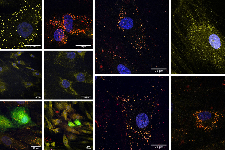
Observation of tumour cells transfected with fluorescently labeled nanoparticles (green) and labelled in their nucleus (blue). Source: doctoral thesis by Dr Laura Balcells.
Confocal microscopy is a widely used tool in biomedical areas, which offers the possibility of obtaining images with high spatial resolution due to the principle of confocality, which allows directing a laser light that illuminates the sample to a focal plane defined by the user.
This microscopic imaging technique consists of acquiring images at different planes for the subsequent three-dimensional reconstruction of the visualized structures. Specifically, it involves the ability to focus on a single spatial plane and centre the light at that point, which enables high resolution fluorescence imaging. In fact, much higher levels of resolution are obtained with the same magnifications that a confocal fluorescence microscope uses.
Confocal microscopy is used for any application that involves the analysis of images that emit fluorescence, on their own or if they have been previously labeled with fluorophores, whether the cells themselves or cell cultures.
IQS has a Leica SP8 Confocal Scanning Microscope equipped with four lasers that emit at different wavelengths and that, once combined, make it possible to view a wide range of fluorophore combinations and observe cell dynamics in detail as well. The equipment also has three objectives with different magnifications, which enables observing samples that need more or less amplification.
Due to its particular characteristics, the Leica SP8 confocal microscope also facilitates the analysis of materials and biomaterials in addition to the analysis of biological tissues and cell culture.
Leica SP8 confocal microscope applications
The confocal microscope is one of the tools used within the different research areas of the IQS School of Engineering Materials Engineering Group (GEMAT), especially in areas of Biomedical research.
Thus, for example, confocal microscopy techniques are used to determine internalization kinetics of nanoparticles within different types of cells, as well as to see their intracellular location. For a specific example, this technique has been used to see how metallic nanoparticles are internalized in tumour cells, different from how they do in healthy cells, and how they manage to produce a modulation in the natural processes of cellular endocytosis and exocytosis.
Other examples of applications for the device can be found in the field of Biomaterials Engineering in which, for example, it is used to observe polymeric coatings of electrosprayed fibres.
This device has been co-financed by the European Regional Development Fund of the European Union within the framework of the ERDF Operational Programme of Catalonia 2014-2020 (file 2015 ERDF S-05).

